A Deep Dive Into How Vegetable Oils Drive Chronic Disease

I have written extensively about the perils of over-consumption of omega-6 oils. You can read some of my posts here, here, here, here, here, here, here, here, here, here, and here. I kept those posts relatively simple. But in this post, I want to focus on the damage they cause at the cellular level, which will make it rich in scientific terms and explanations. This post is intended for those who want to take a deeper dive into the actual mechanisms of damage caused by omega-6 oils.

Few dietary changes in human history have been as profound—or as overlooked—as the explosive rise of omega-6-rich seed oils. Before the late 19th century, human intake of linoleic acid (the dominant omega-6 fatty acid in these oils) hovered below 2 percent of total calories. Today, in Westernized diets, it often exceeds 10 percent, with some individuals reaching 15 percent or more.¹ These oils now supply roughly one-third of daily calories in processed foods and restaurant meals. Dr. Chris Knobbe, founder of the Cure AMD Foundation and co-author of The Ancestral Diet Revolution, has spent years documenting how this shift is not merely correlative but causal at the cellular level. The damage begins with the incorporation of these highly unsaturated fats into our most fundamental structures and spirals into systemic metabolic collapse.² Recognizing this helps you understand the importance of dietary choices for your overall health.
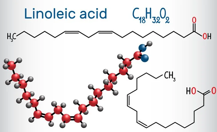
The trouble starts in cell membranes. Omega-6 fatty acids, especially linoleic acid (LA, an 18-carbon chain with two double bonds), are readily absorbed and esterified into phospholipids that form the lipid bilayers of every cell. Because they are polyunsaturated, they introduce multiple cis double bonds that create kinks in the chain. This fluidity is useful in small amounts, but excess LA makes membranes abnormally prone to oxidation. Once embedded, LA and its elongated derivative, arachidonic acid (AA), become substrates for non-enzymatic lipid peroxidation upon encountering reactive oxygen species (ROS) generated during normal metabolism. The result is a self-propagating chain reaction that produces toxic advanced lipid oxidation end-products (ALEs) such as 4-hydroxynonenal (4-HNE), malondialdehyde (MDA), and hydroxyoctadecadienoic acids (HODEs). These aldehydes are cytotoxic, genotoxic, mutagenic, and highly reactive; they cross-link proteins, damage DNA, and trigger inflammatory signaling.³
Nowhere is this peroxidation more devastating than inside the mitochondria—the cellular power plants responsible for generating roughly 90 percent of our ATP. Mitochondrial inner membranes are uniquely rich in cardiolipin, a diphosphatidylglycerol phospholipid that contains four fatty-acid tails, often including linoleic acid in modern diets. Knobbe’s analysis shows that high omega-6 intake directly damages the structure of cardiolipin. Oxidized cardiolipin loses its characteristic curvature and tight packing. The inner membrane becomes “leaky”: protons that should be pumped across the gradient during electron transport escape back into the matrix without driving ATP synthase. Energy production collapses. At the same time, electron leakage from the dysfunctional electron transport chain dramatically increases ROS output. A vicious cycle ensues—more oxidation, more membrane damage, more ROS, less ATP. Cells enter a state of energetic starvation and chronic oxidative stress.⁴
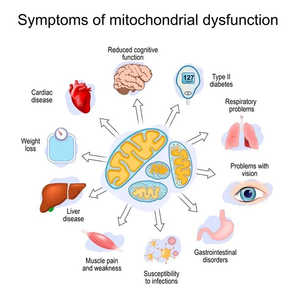
This mitochondrial dysfunction manifests first as cellular insulin resistance. When mitochondria cannot produce adequate ATP or when ROS overwhelm antioxidant defenses, insulin signaling is impaired at the receptor and post-receptor levels. Glucose uptake falters. The cell, desperate for energy, shifts to inefficient glycolysis and fat storage. Scaled across billions of cells, this produces whole-body insulin resistance, hyperinsulinemia, and the metabolic syndrome that Knobbe links to obesity and type 2 diabetes. Notably, animal studies demonstrate that iso-caloric diets differing only in omega-6 content (19 percent soybean oil versus 1 percent LA) produce massive obesity in rodents—equivalent to a 277-pound human versus a 170-pound human—without added sugar or excess calories. The driver is mitochondrial failure, not caloric surplus.⁵
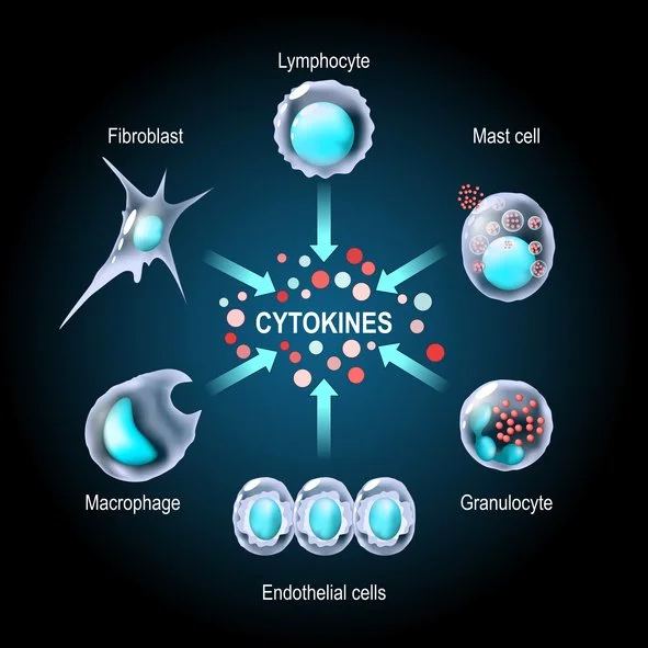
Beyond energy failure, the inflammatory cascade amplifies harm. Arachidonic acid, derived from dietary LA, serves as a substrate for cyclooxygenase and lipoxygenase enzymes, yielding proinflammatory eicosanoids: prostaglandins, leukotrienes, and thromboxanes that promote vasoconstriction, platelet aggregation, and immune cell recruitment. Simultaneously, the ALEs activate NF-κB and other transcription factors, which upregulate inflammatory cytokines. Chronic low-grade inflammation damages the endothelium, promotes foam-cell formation, and accelerates atherosclerosis. Oxidized LDL particles, enriched with peroxidized omega-6 lipids, are preferentially taken up by macrophages in the arterial intima, forming fatty streaks and plaques—the true origin of coronary heart disease according to the oxidized-linoleic-acid hypothesis.⁶
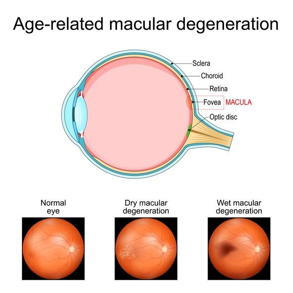
The retina offers a stark window into this pathology. Knobbe’s original research on age-related macular degeneration (AMD) revealed that the disease was virtually unknown before 1850 and remained rare until the 1930s—precisely when seed-oil consumption surged. Photoreceptor outer segments are extraordinarily rich in polyunsaturated fatty acids and exposed to high oxygen tension and light, conditions that accelerate peroxidation. Oxidized lipids accumulate as drusen, destroy retinal pigment epithelium, and kill photoreceptors. The same mechanisms—cardiolipin peroxidation, mitochondrial ROS, and ALE toxicity—explain why AMD, once absent in ancestral populations consuming <2 percent of their calories from omega-6, now blinds millions.⁷
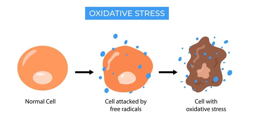
Additional cellular insults compound the picture. Peroxidized lipids trigger ferroptosis, an iron-dependent form of regulated cell death that is particularly lethal in cells with high PUFA content. Genotoxic ALEs form DNA adducts, increasing mutation risk and cancer potential. Adipose tissue itself becomes dysfunctional: omega-6 accumulation enlarges adipocytes, promotes hypoxia, and converts the fat depot from an endocrine organ into a source of systemic inflammation. Nutrient deficiency exacerbates everything; unlike ancestral animal fats rich in vitamins A, D, and K2, seed oils supply none of these fat-soluble cofactors yet displace the very foods that provide them.⁸
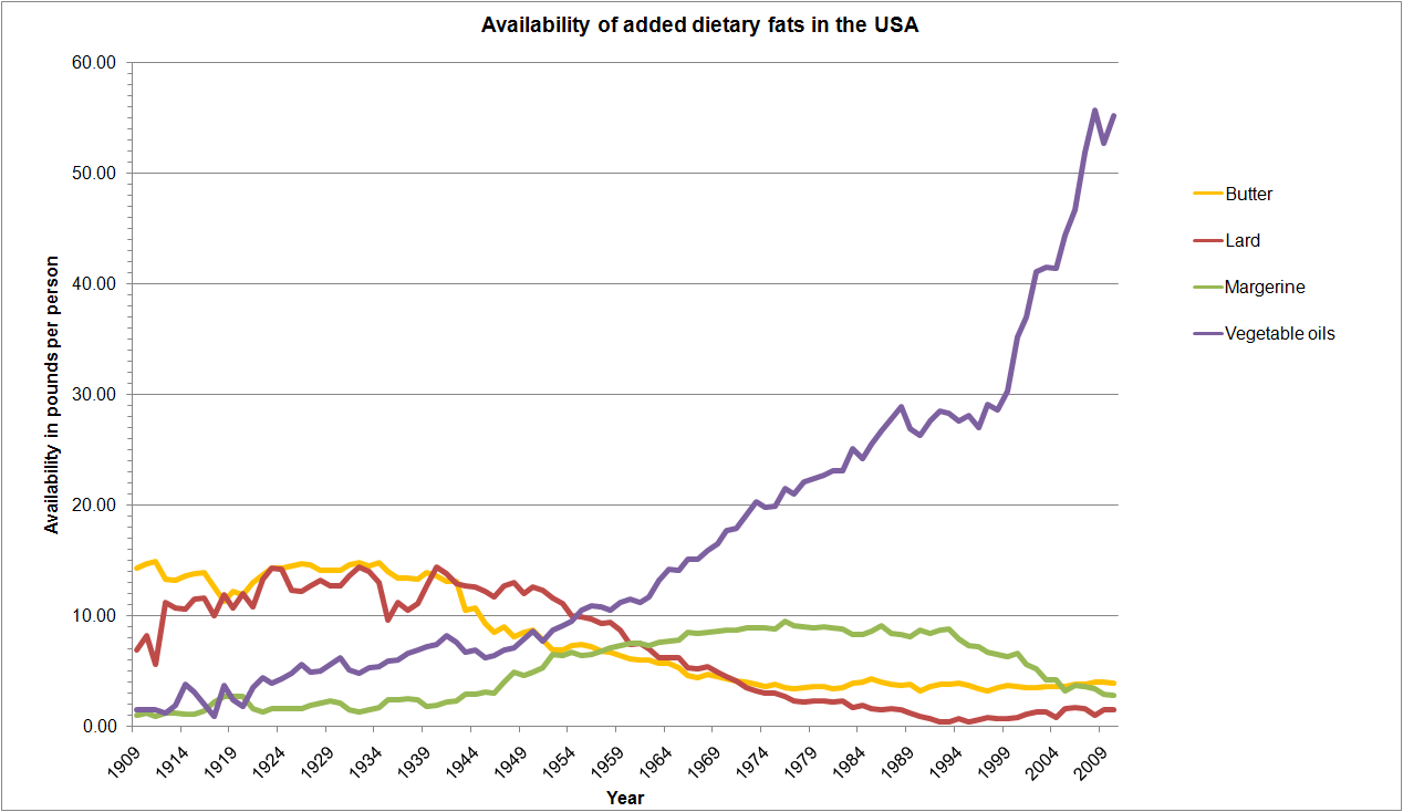
Epidemiological patterns reinforce the cellular story. In the United States, LA in adipose tissue rose from roughly 2.8 percent in ancestral-like populations to 9.1 percent by 1959 and over 21 percent by 2008—mirroring the exponential rise in obesity (from 1.2 percent in 1900 to >40 percent today), coronary heart disease (virtually nonexistent before 1911), and diabetes. Nations that abandoned traditional sources of saturated fat in favor of soybean, canola, and sunflower oils have experienced parallel epidemics. Conversely, populations maintaining <2 percent omega-6 intake show near-zero rates of these “diseases of civilization.”⁹

The cellular damage is cumulative and insidious because fatty-acid turnover in membranes and adipose tissue occurs over years, not days. Once embedded, the pro-oxidative, proinflammatory, and energetically defective state persists until dietary reversal. Knobbe emphasizes that removing seed oils and returning to ancestral fats—tallow, butter, lard, olive oil, and fatty fish—halts the peroxidation cascade, restores mitochondrial integrity, and allows cellular repair. Animal and human data suggest that metabolic recovery can begin within weeks to months, with full membrane remodeling taking up to three years.¹⁰
In summary, diets high in omega-6 seed oils do not merely “promote inflammation” in vague terms; they inflict precise, mechanistic wreckage at the lipid-membrane and mitochondrial levels. Lipid peroxidation generates toxic aldehydes; cardiolipin oxidation impairs ATP synthesis; excess ROS creates a feedback loop of oxidative stress; and downstream eicosanoid and ALE signaling drive insulin resistance, endothelial damage, and chronic disease. Dr. Chris Knobbe’s work, grounded in the historical disappearance of these illnesses, rigorous cross-national correlations, and biochemical plausibility, identifies seed oils as the primary and proximate driver of the modern metabolic catastrophe. The solution is elegantly simple: eliminate the oils that never existed in the human diet until the industrial era and reclaim the fats our cells evolved to use. The cellular evidence is clear—our mitochondria are paying the price every day we ignore it. I will close with my favorite advice: Eat a whole-food diet that includes animal-based foods with their inherent fats, while avoiding processed foods, especially processed carbohydrates and seed oils.
Footnotes
¹ Knobbe CA. Data presented in lectures and summarized in The Ancestral Diet Revolution: How Vegetable Oils and Processed Foods Are Destroying Your Health (2023). Historical consumption figures drawn from USDA economic research and adipose-tissue analyses.
² Knobbe CA, Stojanoska M. The ‘Displacing Foods of Modern Commerce’ Are the Primary and Proximate Cause of Age-Related Macular Degeneration. Medical Hypotheses. 2017;110:23-30. doi:10.1016/j.mehy.2017.10.010.¹
³ Mortensen MS, et al. Polyunsaturated Fatty Acids Drive Lipid Peroxidation and Ferroptosis. Cells. 2023;12(5):804.
⁴ Direct mechanistic description from Knobbe’s interviews and lectures, including damage to cardiolipin, proton leak, and ROS amplification (e.g., Mind & Matter podcast #136, 2023).
⁵ Knobbe CA. Animal feeding studies referenced in public presentations (iso-caloric high- vs. low-LA diets producing differential obesity).
⁶ DiNicolantonio JJ, et al. Omega-6 vegetable oils as a driver of coronary heart disease. Open Heart. 2018;5:e000898.
⁷ Knobbe CA, Stojanoska M. Medical Hypotheses 2017 (op. cit.). Historical AMD rarity documented through global medical literature review 1851–1975.
⁸ Additional pathways including ferroptosis and genotoxicity summarized in lipid-peroxidation literature (Mortensen et al., 2023) and Knobbe’s broader disease model.
⁹ Adipose LA percentages: Pryor et al. (1969) ancestral data vs. modern U.S. surveys; disease prevalence trends from CDC, WHO, and historical medical records cited by Knobbe.
¹⁰ Membrane fatty-acid half-life approximately 600–680 days; full turnover cited by Knobbe as ~3 years.